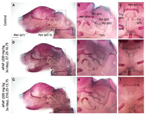
Repeated administration of high APAP dosage to pregnant mice does not recapitulate craniofacial defects. (A–I) Pregnant dams were administered via oral gavage three times daily: Saline between E10.25–13.75 (A–C), 200 mg/kg APAP between E7.25–10.75 (D–F), 200 mg/kg APAP between E10.25–13.75 (G–I). The resulting pups were collected at P0 and stained with alcian blue (cartilage) and alizarin red (bone). (A–C) A control pup with pertinent bones and cartilages labeled. Pharyngeal arch-derived structures are indicated with parentheses (e.g., “p1” = derived from the first pharyngeal arch). (D–F) In a pup whose mother was treated with APAP between E7.25–10.75, all labeled structures develop normally. (G–I) No defects seen in a pup whose mother was treated with APAP between E10.25–13.75. Dashed boxes in (A,D,G) are the fields of view for panels (B,E,H), respectively. Abbreviations: cricoid cartilage, Cri; hyoid, Hyo; incus, Inc; malleus, Mal; mandible, Man; stapes, Sta; styoid process of the temporal bone, Sty; thyroid cartilage, Thy; tracheal cartilages, Tra; tympanic, Tym. Scale bars in (A–C), 1 mm.
|
