
FIGURE 4
- ID
- ZDB-FIG-220723-26
- Publication
- Shin et al., 2021 - Back and forth: History of and new insights on the vertebrate lymphatic valve
- Other Figures
- All Figure Page
- Back to All Figure Page
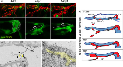
Cellular and ultrastructural features of zebrafish larval lymphatic valves. (a) Confocal images of facial lymphatic vessels at indicated time point. Lymphatic endothelial cells are in red, lymphatic valve (LV) cells also express the gata2a:gfp transgene in green. Bottom panels are magnified region indicated in corresponding top image; only green channel is shown. Valve cell shapes traced with dotted lines at 4 dpf. Lateral view, anterior to left, dorsal is up. (b) Scanning electron microgram of lymphatic valve leaflets (indicated by arrowheads). Mural cells (MCs) adjacent to lymphatic endothelial cells are shown. Yellow box indicates region magnified in (c). (c) Extracellular matrix (ECM) core between two lymphatic valve endothelial cells is pseudocolored in yellow. Fibril-bundles are shown by arrows and electron dense regions suggestive of matrix binding are denoted by arrowheads. (d) Schematic depicting facial lymphatic vessel (red) morphogenesis; primary head sinus (PHS) shown in blue. Lymphatic valve progenitors and valve shown in green. CCV, common cardinal vein; FLS, facial lymphatic sprout; FLV, facial lymphatic vessel; PHS-LPA, primary head sinus-anterior lymphatic progenitor; PHS-LPP, primary head sinus-posterior lymphatic progenitor. Images (a–c) are re-used from Shin et al. (2019) with permission |