
FIGURE 2
- ID
- ZDB-FIG-220723-24
- Publication
- Shin et al., 2021 - Back and forth: History of and new insights on the vertebrate lymphatic valve
- Other Figures
- All Figure Page
- Back to All Figure Page
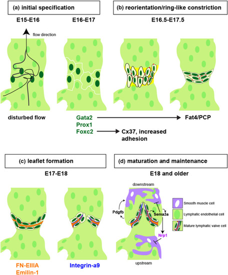
A schematic drawing of lymphatic valve development in collecting lymphatic vessel of mouse model adjusted and modified from (Koltowska et al., 2013; Tatin et al., 2013). (a) From E15~E16, disturbed lymph flow (from bottom to top) downstream from a branch point induces Foxc2 and Gata2 expression, which in turn induces Prox1 to initiate valve development at E16-E17 (Cha et al., 2018; Kazenwadel et al., 2015; Norrmén et al., 2009; Petrova et al., 2004; Qu et al., 2015; Sabine et al., 2012; Zhou et al., 2010). (b) From E16.5 to E17.5, key transcription factors stimulated by shear stress regulate cell-cell adhesion (e.g. Foxc2 and Cx37) and cell polarity (e.g. Gata2 and Fat4) to promote reorganization of lymphatic valve cells to form a ring-like constriction with expression of basement membrane proteins (yellow). (c) Between E17 and E18, LvEC secrete ligands for Integrin-a9 (orange, FN-EIIIA and Emilin-1) into an extracellular matrix core, and express Integrin-a9 on their basal surface (blue lines) to promote valve leaflet extension. (d) At E18.0 or later, lymphatic valves are fully mature and collecting lymphatic vessels exhibit smooth muscle coverage (SMCs, purple). SMCs are recruited by Pdgfb, except at the valve, where they are repelled by Sema3a binding to Nrp1, a direct target of Nrp1. Subcellular types in mature lymphatic valves are color-coded and listed |