
Fig. 9
- ID
- ZDB-FIG-220705-40
- Publication
- Chen et al., 2022 - Photosubstitution in a trisheteroleptic ruthenium complex inhibits conjunctival melanoma growth in a zebrafish orthotopic xenograft model
- Other Figures
- All Figure Page
- Back to All Figure Page
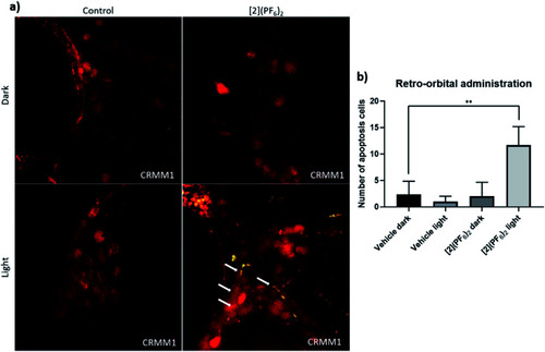
TUNEL assay in the CRMM1 orthotopic tumour model after RO injection of [2](PF6)2. (a) Red fluorescent CRMM1 cells were injected behind the eye of the embryo at 2 dpf, and the embryos were divided into four groups for drug treatment. RO administration of vehicle control and [Fig. 8. After dark or light exposure, embryos were fixed and TUNEL staining was performed. (a) Representative overlay images of embryos are shown. In the group treated with [2](PF6)2 and light, nuclear DNA fragmentation in nucleases is detected by co-localization of green (DNA fragments) and red (CM tumour cell) signal, depicted on the overlay as yellow signal marked by white arrows. In the dark control group, light control group, and group treated with [2](PF6)2 and left in the dark, there were no positive green apoptotic tumour cells. The background green signal in the [2](PF6)2 light groups did not co-localize with cytosolic red signal, which is diminished in degraded cells and TUNEL stains only the DNA breaks in these CM apoptotic cells. (b) Quantification of the number of apoptotic tumour cells (yellow dots). Experiment was performed 3 times with a group size of 10 embryos. **P < 0.01. |