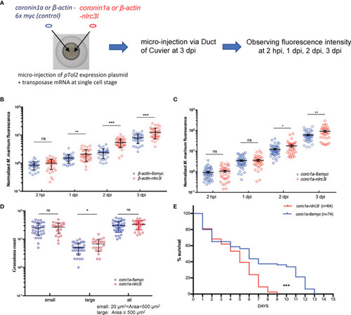
The impact of nlrc3-like on host defense against mycobacterial infection was mainly mediated by innate immune cells. (A) Schematic view of the construction of nlrc3-like overexpressing embryos and the M. marinum infection assay. pTol2 plasmids were injected at the single-cell stage and subsequently the grown embryos were infected via the duct of Cuvier at 3 dpf. Injections of pTol2-β-actin-nlrc3-like (β-actin-nlrc3l) and pTol2-coronin1a-nlrc3-like (coro1a-nlrc3l) generated embryos overexpressing nlrc3-like in all cells or only in myeloid cells, respectively. Black arrows indicate injection sites. (B) Graph of the relative green fluorescence intensity of the β-actin-nlrc3l group and the β-actin-6xmyc group at the indicated time points. (C) Graph of the relative green fluorescence intensity of the coro1a-nlrc3l and coro1a-6xmyc embryos at the indicated time points. (D) Graph showing the number of granuloma-like structures in the coro1a-nlrc3l and the coro1a-6xmyc embryos. (E) Survival curve of coro1a-nlrc3l and coro1a-6xmyc embryos. The data shown represents the averages from two independent biological replicates *p ≤ 0.05; **p ≤ 0.01; ***p ≤ 0.001; ns, not statistically significant (p>0.05).
|
