
FIGURE
Fig. 6
- ID
- ZDB-FIG-220615-33
- Publication
- Yue et al., 2022 - Serotonin (5-HT) 2A Receptor Involvement in Melanin Synthesis and Transfer via Activating the PKA/CREB Signaling Pathway
- Other Figures
- All Figure Page
- Back to All Figure Page
Fig. 6
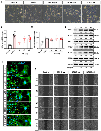
Figure 6. HTR2A agonist DOI induces dendrites and migration of B16F10 cells: (a) The cell morphology of B16F10 cells treated with different concentrations of DOI (10, 20, and 30 μM) and α-MSH (50 nM) is used as a positive control. Scale bar, 50 μm; (b,c) the dendritic number (b) and length (c) of the B16F10 cells treated with different concentrations of DOI (10, 20, and 30 μM) (n = 15); (d) Western blot shows the protein expression of GP100 (melanosome organization), RAB7/17/27 (melanosome transport), RAC1, and CDC42 (melanocytes dendrites) in B16F10 cells treated with DOI (10, 20, and 30 μM); (e) phalloidin staining shows the cytoskeleton in B16F10 cells treated with DOI (10, 20, and 30 μM). DAPI was used to mark the nucleus. The right row is the partial enlargement of the photos on the left. Scale bar, 20 μm (left), 10 μm (right); (f) cell scratch experiments show the migration of B16F10 after treatment with DOI (10, 20, and 30 μM) from 0 to 24 h. Percentage (%) of gap filling/wound healing was used to indicate the migration ability of B16F10 cells. Scale bar, 10 mm. * (p < 0.05), ** (p < 0.01) and *** (p < 0.001) compared to control group.
|
Expression Data
Expression Detail
Antibody Labeling
Phenotype Data
Phenotype Detail
Acknowledgments
This image is the copyrighted work of the attributed author or publisher, and
ZFIN has permission only to display this image to its users.
Additional permissions should be obtained from the applicable author or publisher of the image.
Full text @ Int. J. Mol. Sci.