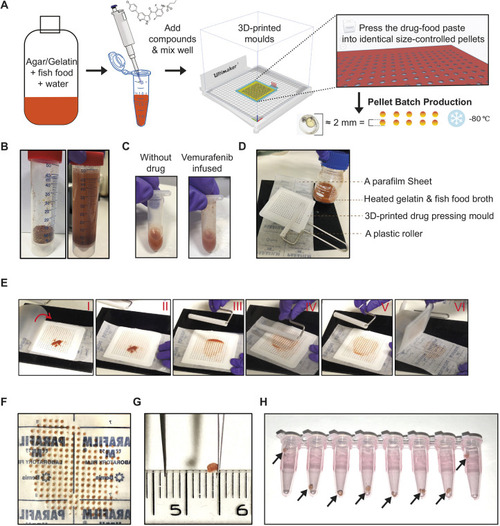
Technical setup and drug pellet preparation. (A) Schematic overview of the drug-pellet formulation and manufacturing pipeline. (B) Dry fish food mix and the food–agar mixture resuspended in water. (C) The red-colour food–agar paste becomes pink once supplemented with vemurafenib dissolved in DMSO. (D) The tools used for pressing drug pellets in the mould. (E) Sequential series of photos (I-VI) showing the process of drug-pellet pressing. The parafilm sheet is peeled from the backing paper, and the mould is placed on the backing paper. The drug paste is applied on to the mould (I), and the parafilm sheet is gently lowered to cover the paste and mould (II). Next, using the roller, the drug paste is evenly applied into the holes of the mould (III-V). The parafilm sheet is lifted, followed by carefully removing the mould, and the drug pellets adhere to the backing paper (VI). (F) A freshly prepared batch of drug pellets. Surface tension retains the pellets on the parafilm backing paper. (G) A drug pellet recovered from −80°C storage, maintaining the flat-cylinder shape. The ruler shown in the picture is scaled in cm/mm. (H) Drug pellets aliquoted into daily doses per fish in PCR tubes, ready for −80°C storage, with arrows highlighting the drug pellets inside the tubes.
|
