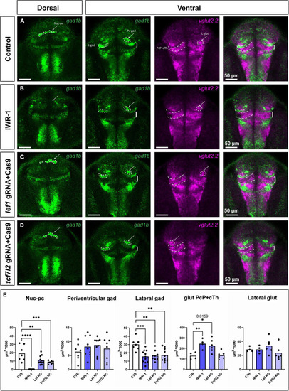
Phenotypes in the pretectum of IWR-1-treated, lef1 crispant and tcf7l2 crispant zebrafish at 48 hpf. Confocal Z-stack images showing brain sections stained using in situ hybridization with gad1b and vglut2.2 probes to visualize different neurochemical clusters of neurons. (A) Control: the gad1b-high Nuc-pc is visible in the dorsal section; gad1b-high lateral and paraventricular clusters in the ventral section; and vglut2.2-positive lateral cluster and vglut2.2-positive region of PcP and cTh are visible in the ventral section. (B) IWR-1: the Nuc-pc is absent, the lateral gad1b-high cluster is smaller, and the vglut2.2-positive area of PcP and cTh is bigger. (C) lef1 crispants: The lateral gad1b-high cluster is smaller, and the vglut2.2-positive area of PcP and cTh is bigger. (D) tcf7l2 crispants: Nuc-pc is smaller and the lateral gad1b-high cluster is smaller. (E) The graphs show volume quantification of distinct clusters indicated in the previous images. Statistics in (E)n = 4–10; one-way ANOVA followed by Dunnett’s post hoc test; *p < 0.05 (the exact p-value is indicated in the graph), **p < 0.01, ***p < 0.001 and ****p < 0.0001. Nuc-pc, nucleus of the posterior commissure; L gad, lateral gad1b-positive cluster; L glut, lateral vglut2.2-positive cluster; Pv gad, periventricular gad1b-positive cluster; CoP, commissural pretectum; JcP, juxtacommissural pretectum; PcP, precommissural pretectum; cTh, caudal thalamus.
|
