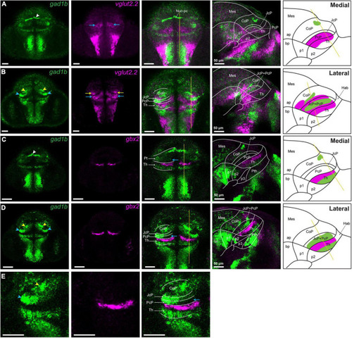
vglut2.2-positive glutamatergic clusters in the pretectum of 48 hpf zebrafish. Confocal Z-stack images showing brain sections stained using in situ hybridization with gad1b,vglut2.2 and gbx2 probes. (A,B) Co-staining of gad1b and vglut2.2 identifies vglut2.2-positive glutamatergic areas in the diencephalon. Low gad1b signal is present throughout the central domain of CoP. (C,D) Co-staining of gad1b and gbx2 identifies the cTh and rTh by the gad1b expression. (E) Magnification of gad1b and gbx2 ventral sections shows the boundary between Pt and Th. White, yellow and blue arrowheads indicate the dorsocaudal, periventricular and lateral gad1b GABAergic clusters, respectively. Yellow and blue arrows indicate vglut2.2 positive glutamatergic clusters in the CoP and PcP, respectively. The schemes depict the regions identified by the markers in each merged sagittal image. ap, alar plate; bp, basal plate; Hab, habenula; Mes, mesencephalon; Nuc-pc, nucleus of posterior commissure; Pt, pretectum; CoP, commissural pretectum; JcP, juxtacommissural pretectum; PcP, precommissural pretectum; p1; prosomere 1; p2, prosomere 2; p3, prosomere 3; Th, thalamus, cTh, caudal thalamus, rTh, rostral thalamus.
|
