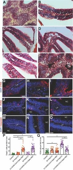
Intranasal delivery of SARS-CoV-2 S RDB protein causes severe cellular changes to the OO of adult zebrafish. (A) H&E image of raphe of the OO showing large eosinophilic deposits 3 d post intranasal delivery of SARS-CoV-2 S RBD (B) Masson Trichrome stains of zebrafish OO administered intranasally vehicle or (C-D) 3 h SARS-CoV-2 S RBD, or (E-F) 3 d SARS-CoV-2 S RBD. Asterisk denotes red keratin deposits. Blue staining corresponds to collagen deposits. (G) anti-CTK8 (red) immunofluorescence staining of adult zebrafish OO cryosections after intranasal delivery of vehicle or (H) 3 h; or (I) 3 d SARS-CoV-2 S RBD. (J) anti-PCNA immunofluorescence staining of adult zebrafish OO cryosection after intranasal delivery of vehicle or (K) 3 h; or (L) 3 d recombinant SARS-CoV-2 S RBD. (M) anti-caspase3 (red) immunofluorescence staining of adult zebrafish OO cryosections after intranasal delivery of vehicle or (N) 3 h; (O) 3 d recombinant SARS-CoV-2 S RBD. Nuclei are stained with DAPI (blue). Scale in (G-O): 20μm. (P) Quantification of the mean number of caspase3 positive cells per field of view. (Q) Quantification of number of PCNA positive cells per field of view. Quantifications shown in P and Q were performed on N = 4 animals and n=10 fields of view per animal, represented by different colors. Error bars represent S.E.M. LP: lamina propria; NC: nasal cavity. Statistics ANOVA followed by a Tukey’s post hoc comparison. *P-value<0.05. Representative images of Poly(I:C)-treated animals are shown in Supplemental figure 2.
|
