
Fig. 6
- ID
- ZDB-FIG-220308-14
- Publication
- Braunstein et al., 2021 - Basal epidermis collective migration and local sonic hedgehog signaling promote skeletal branching morphogenesis in zebrafish fins
- Other Figures
- All Figure Page
- Back to All Figure Page
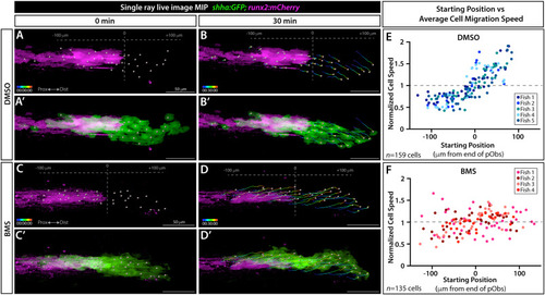
Shh/Smo signaling slows collective migration of shha-expressing basal epidermal cells associated with pre-osteoblasts. (A-D’) Frames from a time lapse movie showing dorsal ray 3 of the caudal fin from live mounted 24 dpf shha:GFP;runx2:mCherry fish treated with DMSO or BMS-833923 (BMS) for 24 h prior to imaging. Images are maximum intensity projections (MIP) and show the start (0 min; A, A’, C, C’) and end (30 min; B, B’, D, D’) points. The Imaris-generated colored tracks show the progressive displacement of individual shha:GFP+basal epidermal cells (green). Grey spheres show the starting or final positions of all tracked basal epidermal cells. Grey dashed vertical lines in (A, B, C, D) indicate the distal most Runx2+ pre-osteoblast (magenta), defined as position “0”. (E, F) Scatter plot graphs showing the average speed of individual basal epidermal cells, considering their net X-displacement and normalized to all scored cells of the given fish, relative to starting position for DMSO- (159 individual cells from five fish) and BMS-exposed fish (135 cells from four fish). Dot colors correspond to cells from a given fish. Scale bars are 50 μm. |
Reprinted from Developmental Biology, 477, Braunstein, J.A., Robbins, A.E., Stewart, S., Stankunas, K., Basal epidermis collective migration and local sonic hedgehog signaling promote skeletal branching morphogenesis in zebrafish fins, 177-190, Copyright (2021) with permission from Elsevier. Full text @ Dev. Biol.