
Fig. 5
- ID
- ZDB-FIG-220308-13
- Publication
- Braunstein et al., 2021 - Basal epidermis collective migration and local sonic hedgehog signaling promote skeletal branching morphogenesis in zebrafish fins
- Other Figures
- All Figure Page
- Back to All Figure Page
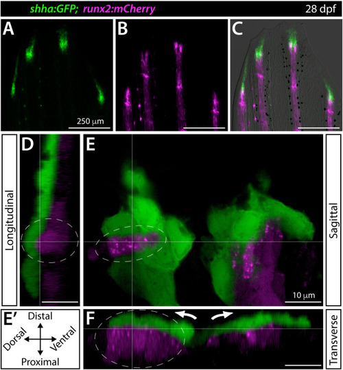
Shha+basal epidermis and pre-osteoblasts are intertwined in developing fins. (A–F) Fluorescence widefield (A–C) or confocal (D–F) images of the dorsal caudal fin lobe of a 28 dpf shha:GFP;runx2:mCherry fish. (A–C) shha:GFP-expressing basal epidermal cells (green) overlay and extend distally from runx:mCherry-high pre-osteoblast (magenta). The overlay in (C) includes a brightfield image for context. (D–F) A single optical slice (E; sagittal; orientation key in E’) and reconstructed longitudinal (D) and transverse (F) views of a distal ray region undergoing ray branching. shha:GFP+basal epidermis (green) and runx2:mCherry+pre-osteoblasts (magenta) have overlapping signal at interfaces, indicating their tight juxtaposition. Basal epidermis and pre-osteoblasts tandemly separate into split pools during branching (white arrows). The grey dotted oval highlights a ridge of pre-osteoblasts nestled into a shha:GFP+basal epidermal groove (Movie 2). Scale bars are 250 μm (A–C) and 10 μm (D–F). |
Reprinted from Developmental Biology, 477, Braunstein, J.A., Robbins, A.E., Stewart, S., Stankunas, K., Basal epidermis collective migration and local sonic hedgehog signaling promote skeletal branching morphogenesis in zebrafish fins, 177-190, Copyright (2021) with permission from Elsevier. Full text @ Dev. Biol.