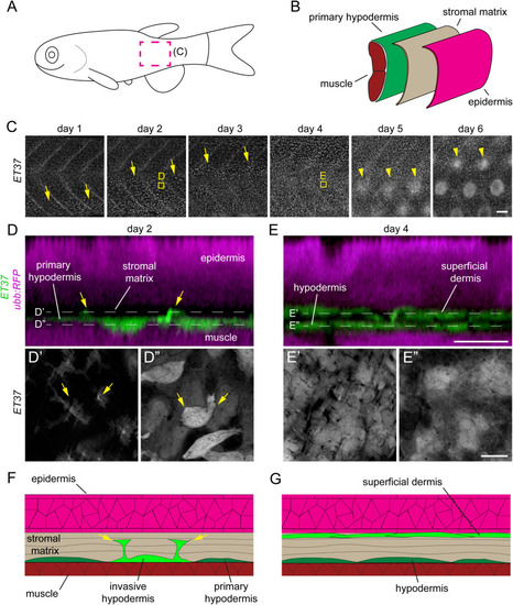
Postembryonic dermal morphogenesis involves superficial directed migration of primary hypodermal cells. (A) Schematic representation of a larval zebrafish with imaged region (C) marked by magenta dotted line. (B) Schematic coronal section of zebrafish trunk showing relative location of the primary hypodermis (green), collagenous stroma (beige) and epidermis (magenta). (C) Time series of late larval dermal morphogenesis visualized in ET37:EGFP; ubb:RFP transgenics. RFP channel omitted for clarity. On day 1 of imaging the first migratory dermal cells appeared near the ventral side of the skin as rounded cells with relatively intense ET37:EGFP expression (yellow arrows). Over the following three days, dermal migration swept dorsally and was completed by day 4. Scale papillae (yellow arrowheads) developed after dermal stratification had completed and followed a similar ventral–dorsal wave. Images are from a single representative individual (n = 8 fish total). (D,E) Super-resolution imaging of boxed regions in C, showing early (B) and completed (C) dermal stratification. Initially (day 2), the subset of primary hypodermal that express elevated ET37:EGFP extended (yellow arrows) toward the epidermis (labelled with ubb:RFP). Two days later (day 4) the dermis had been remodeled into a multilayer tissue. (F,G) Schematic representation of images in (D,E) showing early (F) and completed (G) dermal stratification. Thickness of collagenous stromal matrix is exaggerated for clarity.
|
