|
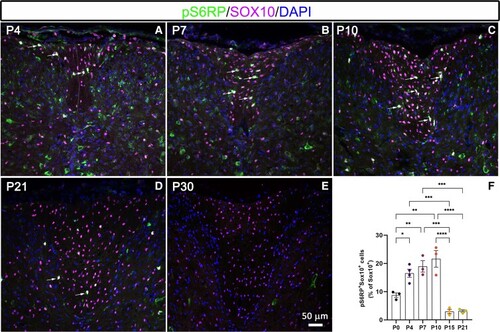
Developmental expression profile of pS6RP in oligodendroglial cells. (A–E) Co-immunolabelling of pS6RP and Sox10 at PND 4 (A), PND 7 (B), PND 10 (C), PND 21 (D) and PND 30 (E) in the spinal cord. Double-positive cells are indicated by arrows. (F) Quantification of pS6RP+Sox10+ cells shows a peak of pS6RP expression between PND7 and PND10, indicating that pS6RP expression is regulated in oligodendroglial cells during development. Data represent mean ± SEM; N = 3 independent experiments for each developmental stage. ANOVA followed with post hoc Tukey's pairwise multiple comparison tests: *P < 0.05; **P < 0.01; ***P ≤ 0.001; ****P ≤ 0.0001. Panels (A–E) are counterstained with DAPI. Scale bar (A–E): 50 µm.
|