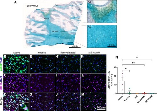
Immunodetection of pS6RP in MS lesions. (A) Luxol fast blue/MHCII staining illustrating an active MS lesion located in the cerebellar white matter. The boxed areas in panel A illustrate a typical active plaque filled with MHCII+ immune cells (A1) and the normal-appearing white matter (A2), respectively. Co-immunolabelling for pS6RP (green) and Sox10 (magenta) indicates that pS6RP is mainly detected in oligodendroglia (arrows) in active (B–D, inset in D) but not in chronic inactive (E–G) nor in remyelinated lesions (H–J) and normal-appearing white matter (K–M). Quantification of the percentage of Sox10+ oligodendroglial cells expressing pS6RP in different MS lesions subtypes and normal-appearing white matter from MS and controls cases (N). Data represent mean ± SEM for each MS lesion type. Active: active lesions (n = 3) and rims of chronic active lesions (n = 1); Inactive: Chronic inactive (n = 3) and core of chronic active (n = 1) lesions; RM: remyelinated lesions (n = 2); MS NAWM: normal-appearing white matter from MS cases (n = 4); Ctl NAWM: normal-appearing white matter from controls (n = 3). ANOVA followed with post hoc Tukey's pairwise multiple comparison tests: *P ≤ 0.05; **P ≤ 0.01. Panels (B–M) are counterstained with Dapi. Scale bars: (A), 5 mm; (A1 and A2), 1 mm; (B–M), 100 µm.
|
