
Figure 1
- ID
- ZDB-FIG-220302-117
- Publication
- Xie et al., 2022 - Two Types of Liposomal Formulations Improve the Therapeutic Ratio of Prednisolone Phosphate in a Zebrafish Model for Inflammation
- Other Figures
- All Figure Page
- Back to All Figure Page
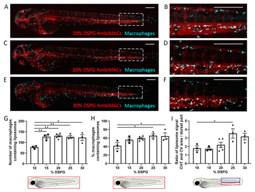
Biodistribution of AmbiMACs with different formulations in zebrafish embryos. (A–F) Representative images of Tg(mpeg1:GFP) embryos injected with AmbiMACs containing different percentages of DSPG at 2 days post-fertilization (dpf). Confocal microscopy images were taken at 2 h post-injection (hpi). AmbiMACs are shown in red and macrophages in cyan. The tail regions (indicated by the dashed boxes in (A,C,E)) are shown at higher magnification in (B,D,F). Scale bar = 200 μm. (G,H) The number (G) and percentage (H) of macrophages containing AmbiMACs quantified in the whole body. A significant difference was observed for the number of macrophages containing AmbiMACs with DSPG percentages of 15–30% compared to 10%. For the percentage of macrophages containing liposomes, a significant difference was observed for AmbiMACs with 25% and 30% DSPG compared to the 10% DSPG. (I) The ratio between the (fluorescent) signal of AmbiMACs in the area, indicated by the red box, encompassing the caudal vein (CV) and the caudal hematopoietic tissue (CHT), and the signal in the dorsal part of the tail (indicated by the blue box). A significant difference was observed between injection with AmbiMACs (25% DSPG) compared to AmbiMACs (10% DSPG). Statistical analysis was performed by one-way ANOVA with Bonferroni’s post hoc test. Data shown are the mean ± SEM of 3–5 individual embryos, of which the individual data are indicated. Statistically significant differences between groups are indicated by: * p < 0.05; ** p < 0.01. |