
Figure 1
- ID
- ZDB-FIG-220219-110
- Publication
- Sanchez-Gonzalez et al., 2022 - Innate Immune Pathways Promote Oligodendrocyte Progenitor Cell Recruitment to the Injury Site in Adult Zebrafish Brain
- Other Figures
- All Figure Page
- Back to All Figure Page
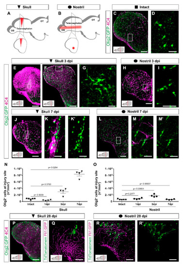
Distinct injury paradigms in the zebrafish telencephalon led to either scarless regeneration or prolonged glial reactivity. (A,B) Schemes depicting skull (A) and nostril (B) injury paradigms. Red triangle (A) and red line (B) illustrate the injury track. (C,D) Micrographs of a telencephalic section showing the distribution of Olig2:GFP+ oligodendroglia and 4C4+ microglia/monocytes in the intact brain. (E,F) Images of 3 dpi skull-injured sections (4C4+ and Olig2:GFP+ cells) at the level of the injury core delineated by a white line (E) and lateral to the injury core depicting the first signs of Olig2:GFP+ cells to accumulation indicated by the boxed area (F). (H) Image showing the distribution of Olig2:GFP+ and 4C4+ cells at 3 dpi after a nostril injury. (G,I) are magnifications of the boxed areas in (F) and (H), depicting Olig2:GFP+ cell distribution. (J–M′) Images showing the reactivity of 4C4+ and Olig2:GFP+ cells at 7 days after skull (J) and nostril (L) injury. (K,K′,M,M′) are magnifications of the boxed area in the respective images. (N,O) Graphs depicting the density of Olig2:GFP+ cells at the injury site after skull (N) and nostril (O) injury. Data are shown as mean ± SEM; each data point represents one animal. Statistical analysis is based on a non-parametric Kruskal–Wallis Test (p-value = 0.0021) with a post-hoc Dunn test (Many-to-One) in N and a one-way ANOVA (p-value = 2.483 × 10⁻⁵) with a post-hoc Dunnett test (many-to-one) in (O). (P) The accumulation of 4C4+ and Olig2:GFP+ cells resolved at 28 days after skull injury. (Q–R′) Images showing the morphology of ependymoglial cells (labelled by electroporation of TdTomatomem) 28 days after skull (Q) and nostril injury (R,R′). While we observed the restoration of the radial morphology of the labelled ependymoglia that contacts the basement membrane after nostril injury (similar to the intact brain), the ependymoglia after skull injury failed to restore radial morphology and built extensive contacts with Fli1-positive blood vessels. All images are full z-projections of a confocal stack; insets indicate the rostro-caudal levels of the sections. Scale bars in (C,E,F,H,J,L,P,Q,R,R′) = 100 µm; Scale bars in (D,G,I,K,K′,M,M′) = 20 µm. Abbreviations: OB: olfactory bulb, OT: optic tectum, dpi: days post-injury; AFOG: acid fuchsin orange G. Symbol description: black triangle: skull injury; black circle: nostril injury. |