FIGURE

Figure 7.

ID
ZDB-FIG-211216-63
Publication
Alejevski et al., 2021 - Conserved role of the urotensin II receptor 4 signalling pathway to control body straightness in a tetrapod
Other Figures
All Figure Page
Back to All Figure Page
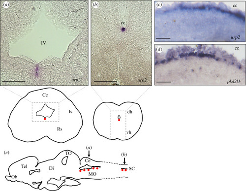
Figure 7.

Localization of urp2-L mRNA in the brain and spinal cord of juvenile X. laevis frog revealed by in situ hybridization. Transverse sections (40 µm) of juvenile frog hindbrain (a) and spinal cord (b) hybridized with antisense urp2-L probe. (c,d) Sagittal sections (40 µm) of juvenile frog spinal cord hybridized with antisense urp2-L (c) and pkd2l1 (d) probes. cc, central canal; IV, fourth ventricle. (e) Schematic sagittal section depicting the distribution of urp2 mRNA hybridization signals (red dots). The level of the coronal sections in (a) and (b) is indicated by arrows. Ce, cerebellum; dh, dorsal horn of the spinal cord; Di, diencephalon; Is, isthmic nucleus; MO, medulla oblongata; Ob, olfactive bulbs; Rs, nucleus reticularis superior; SC, spinal cord; Tel, telencephalon; TO, tectum opticum; vh, ventral horn of the spinal cord. Anatomical structures are designated according to [37]. Scale bars: 150 µm (a,b), 50 µm (c,d).

Expression Data

Expression Detail
Antibody Labeling
Phenotype Data

Phenotype Detail
Acknowledgments
This image is the copyrighted work of the attributed author or publisher, and ZFIN has permission only to display this image to its users. Additional permissions should be obtained from the applicable author or publisher of the image. Full text @ Open Biol.