
Fig. 2
- ID
- ZDB-FIG-211215-2
- Publication
- Pero et al., 2021 - Pathogenic role of delta 2 tubulin in bortezomib-induced peripheral neuropathy
- Other Figures
- All Figure Page
- Back to All Figure Page
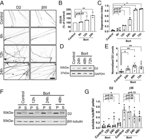
Bort induces D2 at the onset of axonal degeneration. (A) Representative IF images of D2 and βIII staining in axons of DRG neurons (12 DIV) treated with 100 nM of Bort for the indicated times. Scale bar, 50 μm. (B) Ratio analysis of D2/βIII levels measured by IF in axons from fixed neurons treated as in A. Data are pooled from three to four experiments (n = 10 to 41 neurites per condition for each experiment). (C) Time-dependent increase of axonopathy in DRG neurons treated with 100 nM of Bort for the indicated times. Data are from three to four experiments (n = 4 to 15 fields per condition for each experiment). (D) Immunoblot analyses of D2 levels from whole cell lysates of adult DRG neurons (12 DIV) treated with increasing doses of Bort for the indicated times. (E) Quantification of normalized D2 levels as in D. Data are pooled from five to six experiments. (F) Immunoblot analyses of D2 and βIII levels present in the MT pellet (P) and soluble tubulin (S) fractions isolated from adult DRG neurons (12 DIV) and treated with 100 nM of Bort for the indicated times. (G) Quantification of D2 and βIII levels as in F. Data are from four to eight experiments. All data in B, C, E, and G are shown as medians plus interquartile range, and statistics were analyzed by Mann–Whitney U test. *P < 0.05, **P < 0.01. |