
Figure 1.
- ID
- ZDB-FIG-211207-90
- Publication
- Kenney et al., 2021 - A 3D adult zebrafish brain atlas (AZBA) for the digital age
- Other Figures
- All Figure Page
- Back to All Figure Page
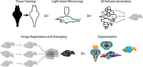
Dissected brain samples were first subject to staining and tissue clearing. This was followed by whole-mount imaging using light-sheet fluorescence microscopy. Three-dimensional volumes were created from individual image sets, and then registered into the same anatomical space prior to averaging to generate a representative image. Finally, volumes were segmented into over 200 neuroanatomical regions.
|