
Figure 4
- ID
- ZDB-FIG-211207-100
- Publication
- Nakajima et al., 2021 - Regenerative Polarity of the Fin Ray in Zebrafish Caudal Fin and Related Tissue Formation on the Cut Surface
- Other Figures
- All Figure Page
- Back to All Figure Page
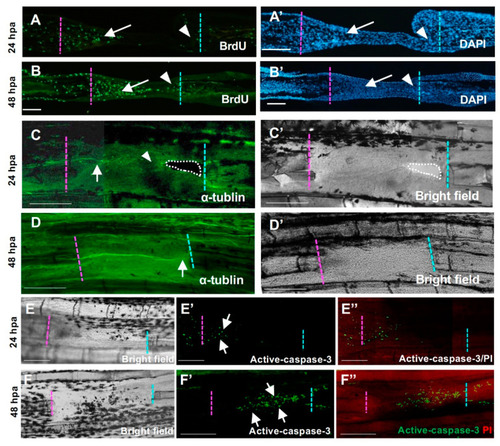
Cellular responses during hole regeneration. (A,A’,B,B’) BrdU-positive cells during hole regeneration. (A) At 24 h post-amputation (hpa), high proliferation of mesenchymal cells was observed in the proximal-derived tissue (arrow), but few cells were observed in the distal side (arrowhead). (B) At 48 hpa, high proliferation of mesenchymal cells was maintained, and proliferation of epithelial cells was also detected. In the distal tissue, some cells were also BrdU-positive (arrowhead). (A’) and (B’) are DAPI staining of (A,B), respectively. Scale bars = 100 μm. (C,C’,D,D’) Nerve distribution during hole regeneration. (C,C’) 24 hpa. (C′) is the bright field view of (C). The nerve fiber extended from the proximal end to the regenerative tissue (arrow). By contrast, few nerve fibers were detected in the apical half of the epithelial sheet and the distal cut end (arrowhead). (D,D’) 48 hpa. (D’) is the bright field view of (D). Several nerve fibers extended from the proximal cut end to the distal cut end (arrow). Scale bars = 300 μm. (E,E’,F,F’) Apoptosis during hole regeneration. (E,F) Bright field. (E’,F’) Active caspase-3. (E”,F”) Merged images of active caspase-3 and propidium iodide (PI) staining. (E,E’,E”) At 24 hpa, apoptosis was only observed in the epithelium covering the blastema formed on the surface of the proximal side (arrow, n = 3). (F,F’,F”) At 48 hpa, apoptotic cells were observed in the tissue derived from the proximal surface (arrow, n = 3). Resection edges (dotted lines). Scale bars = 100 μm (A,B), 300 μm (C,D). |