
Figure 1
- ID
- ZDB-FIG-211201-192
- Publication
- Raby et al., 2021 - Loss of Polycomb Repressive Complex 2 Function Alters Digestive Organ Homeostasis and Neuronal Differentiation in Zebrafish
- Other Figures
- All Figure Page
- Back to All Figure Page
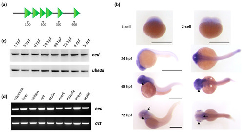
Organization of the Eed protein and eed mRNA expression in zebrafish: (a) schematic representation of the Eed protein. The zebrafish protein, as its human ortholog, is composed of 441 amino acids and contains six WD40 domains (SMART: SM000320) shown as green triangles; (b) whole-mount RNA in situ hybridization showing maternally provided eed transcripts at the 1-cell and 2-cell stage, and zygotic eed mRNA distribution at 24, 48 and 72 hpf. At these later stages, a lateral view is at the left and a dorsal view at the right. The arrowheads show the retina, the white asterisks the pectoral fin buds, and the arrow identify the midbrain-hindbrain boundary. Scale bar is 500 µm; (c) RT-PCR experiment showing the detection of eed transcripts at 1 hpf, 3 hpf, 6 hpf, 24 hpf, 48 hpf, 72 hpf, 4 dpf, and 5 dpf. Ube2a is used as a control; (d) RT-PCR experiment showing eed mRNAs in adult zebrafish tissues. Beta-actin (act) is used as a control. (See Supplementary Materials). |
| Genes: | |
|---|---|
| Fish: | |
| Anatomical Terms: | |
| Stage Range: | 2-cell to Adult |