
Fig. 2
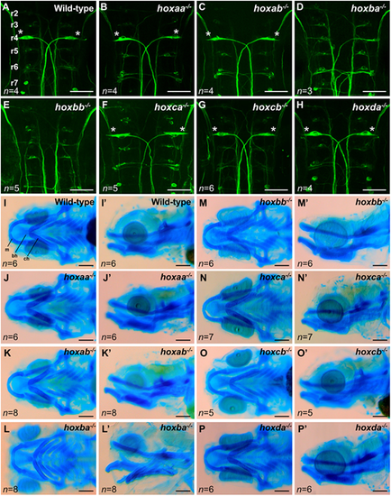
Reticulospinal neurons and craniofacial cartilage in zebrafishhoxcluster mutants. (A-H) Reticulospinal neurons in the hindbrain at 48 hpf were visualized by whole-mount immunostaining using anti-neurofilament RMO-44 antibody. Dorsal view of flat-mounted specimens after removal of the yolk. Mauthner cells located at r4 are indicated by asterisks. Hemizygous mutants for each hox cluster appeared indistinguishable from the wild-type. (I-P′) Craniofacial cartilages of zebrafish larvae at 5 dpf were stained with Alcian Blue. I-P show ventral view and I′-P′ show lateral view. bh, basihyal; ch, ceratohyal; m, Meckel's cartilage. Scale bars, 50 µm (A-H); 100 µm (I-P). |