
FIGURE
Fig. 6
Fig. 6
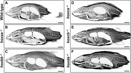
Whole-body tissues ofhoxcluster mutants. (A-F) Whole-body tissues were analyzed by micro-CT scanning. After the micro-CT scanning for skeleton analysis, soft tissues of the same specimens were stained with Lugol's solution, and the stained tissues were subject to micro-CT scanning. Adult males (hoxbb−/− and hoxda−/−) and females (wild-type, hoxaa−/−, hoxca−/− and hoxcb−/−) were used. Eggs are seen in the abdomen of female fish. Movies 11-20 show micro-CT scan 3D movies of transverse and sagittal sections. Scale bars: 2 mm. |
Expression Data
Expression Detail
Antibody Labeling
Phenotype Data
Phenotype Detail
Acknowledgments
This image is the copyrighted work of the attributed author or publisher, and
ZFIN has permission only to display this image to its users.
Additional permissions should be obtained from the applicable author or publisher of the image.
Full text @ Development