
FIGURE
Fig. 3
- ID
- ZDB-FIG-211129-92
- Publication
- Lin et al., 2021 - Two-photon scanned light sheet fluorescence microscopy with axicon imaging for fast volumetric imaging
- Other Figures
- All Figure Page
- Back to All Figure Page
Fig. 3
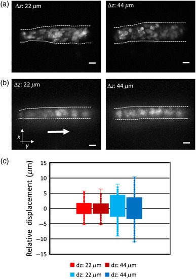
The maximum intensity projection of a volume image (10 slices) of RBCs in Tg(fli1a:EGFP; gata1:DsRed) zebrafish larva over two volume thicknesses using 2p LSFM (a) without and (b) with axicon imaging. (c) Boxplot of RBCs displacement in |
Expression Data
Expression Detail
Antibody Labeling
Phenotype Data
Phenotype Detail
Acknowledgments
This image is the copyrighted work of the attributed author or publisher, and
ZFIN has permission only to display this image to its users.
Additional permissions should be obtained from the applicable author or publisher of the image.
Full text @ J. Biomed. Opt.