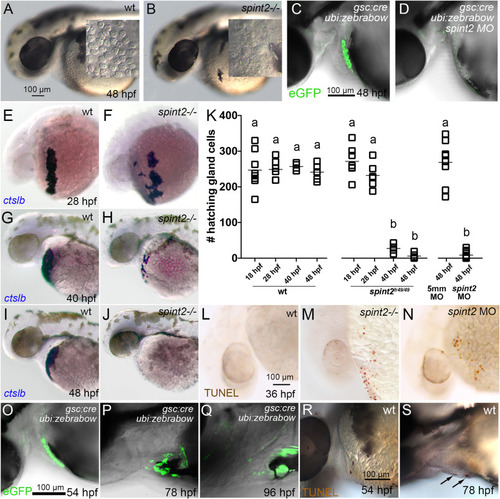
A-B. DIC images of the head and yolk region of 48 hpf old wild-type (A) and spint2fr49/fr49 mutant (B) embryos. Inlets show a magnified view of the hatching gland cell region, in which granule-containing cells are observed in wild-type but not in mutant embryos. C-D. Overlay of DIC and fluorescent images of 48 hpf embryos transgenic for Tg(gsc:Cre)fr44Tg and Tg(ubi:Zebrabow-M)a131. In wild type (C) gfp-expressing hatching gland cells are visible, whereas they are absent in spint2 morphant (D). E-K. WISH of ctslb in wild-type siblings (E,G,I) and spint2 mutants (F,H,J), revealing similar numbers of hatching gland cells in mutants and wild-type siblings at 28 hpf, but progressively fewer hatching gland cells in mutants compared to wild types at 40 hpf and 48 hpf. K. Quantification of numbers of ctslb-positive hatching gland cells at 18 hpf, 28 hpf, 40 hpf, 48 hpf in wild-type and spint2 mutant embryos, and at 48 hpf in spint2 morphants and siblings injected with 5-mismatch (5 mm) control MO. Squares represent single embryos; significances were determined via a one-way ANOVA and Tukey’s post hoc test. Different letters indicate statistically significant differences (p < 0.0001). L-N. TUNEL staining of wild-type (L), spint2 mutant (M) and spint2 morphant (N) embryos at 36 hpf. spint2-deficient embryos display cell death in the hatching gland cells region. O–S. Wild-type hatching gland cells die after hatching. O-Q. Overlays of DIC and fluorescent images of embryos transgenic for Tg(gsc:Cre)fr44Tg and Tg(ubi:Zebrabow-M)a131, showing GFP-positive hatching gland cells shortly after embryos have hatched at 54 hpf (O) but their subsequent loss over the next two days (P,Q). R,S. TUNEL staining of wild-type embryos showing no positive TUNEL cells in the hatching gland tissue in a 54 hpf embryo (R) but few TUNEL-positive hatching gland cells at 78 hpf (S).
|
