
Fig. s1
- ID
- ZDB-FIG-211105-20
- Publication
- Eom et al., 2021 - Immunoglobulin superfamily receptor junctional adhesion molecule 3 (Jam3) requirement for melanophore survival and patterning during formation of zebrafish stripes
- Other Figures
- All Figure Page
- Back to All Figure Page
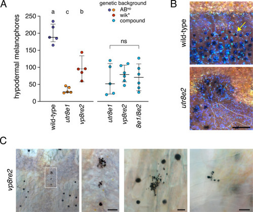
Pigmentary phenotypes of jam3b mutant adults. (A) Numbers of hypodermal melanophores along five segments at the level of the ventral primary melanophore stripe of wild-type and dorsal to the anal fin. Left side of plot indicates melanophores in wild-type and homozygous psrutr8e1 or psrvp8re2 mutants, as maintained in different genetic backgrounds. Each point represents an individual fish and bars show means ± 95% confidence intervals. Different letters indicate means significantly different in post hoc Tukey-Kramer comparisons (all P<0.005; overall ANOVA: F2,12=57.6, P<0.0001). Right side of plot shows lack of significant differences among allelic combinations in a common genetic background (F2,14=0.3, P=0.7488). (B) Details of iridophores typical of interstripes (yellow-gold iridescent cells) and stripes (blue iridescent cells) in both wild-type and psr mutants. Although blue stripe iridophores normally require melanophores for their development (Gur et al., 2020), patches of such iridophores that lacked associated melanophores were often evident in psr mutants (dashed line). Note that melanophore cell bodies and processes extend beyond regions of visible melanin (e.g., arrow) (Hamada et al., 2014), which concentrates towards cell centers during normal physiological modulation of melanosome dispersion and when fish are treated with epinephrine to contract melanosomes from the periphery (Fujii, 2000). Yellow–orange xanthophores are faintly visible over interstripe iridophores. (C) Melanized cell fragments in adult psrvp8re2 mutants. Left two panels, low magnification and detail of melanized debris in hypodermis. Right panels show additional examples in hypodermis and at the surface of a scale, typical of debris being extruded through the epidermis (Lang et al., 2009). Scale bars, 200 μM (B), 50 μM (C). |
Reprinted from Developmental Biology, 476, Eom, D.S., Patterson, L.B., Bostic, R.R., Parichy, D.M., Immunoglobulin superfamily receptor junctional adhesion molecule 3 (Jam3) requirement for melanophore survival and patterning during formation of zebrafish stripes, 314-327, Copyright (2021) with permission from Elsevier. Full text @ Dev. Biol.