
Fig. 7
- ID
- ZDB-FIG-211105-13
- Publication
- Tsedeke et al., 2021 - Cardiomyocyte heterogeneity during zebrafish development and regeneration
- Other Figures
- All Figure Page
- Back to All Figure Page
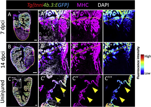
Tg(tnni4b.3:EGFP) expression is downregulated in border zone cardiomyocytes during adult zebrafish heart regeneration. (A, B and C) Sagittal sections from 7 dpci (A), 14 dpci (B) and uninjured (C) Tg(tnni4b.3:EGFP) hearts showing endogenous EGFP expression fluorescence intensity (rainbow gradient), MHC (magenta), and DNA (white). Yellow dotted lines demarcate the injured area. (A′-A‴, B′-B‴) Insets of (A) and (B), respecitvely, showing high-magnification images of MHC+/tnni4b.3:EGFP+ border zone CMs. Yellow dotted lines demarcate the injured area. (C′-C‴) Insets of (C) showing high-magnification images of MHC+/tnni4b.3:EGFP+ AV boundary CMs. Yellow arrowheads point to AV boundary CMs with lower tnni4b.3:EGFP expression. Scale bars: 200 μm (A, B), 100 μm (C). |
Reprinted from Developmental Biology, 476, Tsedeke, A.T., Allanki, S., Gentile, A., Jimenez-Amilburu, V., Rasouli, S.J., Guenther, S., Lai, S.L., Stainier, D.Y.R., Marín-Juez, R., Cardiomyocyte heterogeneity during zebrafish development and regeneration, 259-271, Copyright (2021) with permission from Elsevier. Full text @ Dev. Biol.