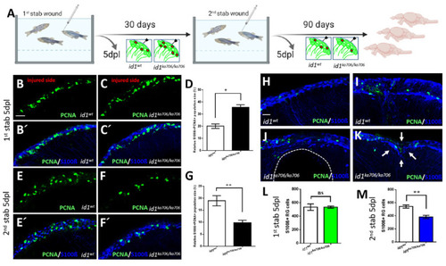
id1 is required to maintain the stem cell pool and the regenerative capacity of the telencephalon. (A) Experimental layout: 6-month-old adult id1ka706/ka706 fish (n = 25) and WT siblings (n = 25) were stabbed with a needle into the right hemisphere of the telencephalon. Fish (n = 2) of each group were sacrificed at 5 days post lesion (dpl) and the remaining fish were allowed to recover for one month before a second stab wound was inflicted. Five days after the second lesion, fish (n = 7) from each group were sacrificed for analysis. The remaining fish were kept for another 3 months before they were sacrificed (n = 16 for WT and n = 11 for id1ka706/ka706 mutants). Note that five mutant fish showed signs of suffering after the second wounding and were sacrificed before the end of the three months recovery. (B–C’, E–F’, H–K) Double immunohistochemistry with S100β (blue) and PCNA (green) antibodies was carried out to mark proliferating NSCs on telencephalic cross-sections from id1ka706/ka706 mutants and WT siblings at the different time points. (B–C’) After the first stab, id1ka706/ka706 mutants showed a higher number of proliferating NSCs. (D) Quantification of the population size of PCNA+/S100β+ cells in relation to the total number of NSCs (S100β+ cells) in WT and id1ka706/ka706 telencephalic cross-sections after the first stab wound. (E–F’) After the second injury, id1ka706/706 mutants showed less proliferating NSCs (PCNA+/S100β+ cells) compared to WT siblings. (G) Quantification of the population size of PCNA+/S100β+ cells in relation to the total number of NSCs (S100β+ cells) in cross-sections through WT and id1ka706/ka706 telencephala. (H–K) WT fish had repaired the lesion in the telencephalon 3 months after the second stab without signs of the injury (H) or with only mild signs of slight tissue disorganization (I). In contrast, id1ka706/ka706 mutants showed severe tissue lesions (J), such as a hole in the parenchyma of the telencephalon (dashed line) or dents in the parenchyma with tissue disorganization (K, white arrows). (L,M) Quantification of the NSCs (S100β+ cells) in the injured side after inflicting the first (L) and second (M) stab wound in WT and id1ka706/ka706 mutants showing a stepwise reduction of the number of NSCs. (Compare also with the increased number of NSCs in the uninjured mutant (Figure S3C)). Significance is indicated by asterisks: ns, not significant; * 0.01≤ p < 0.05; ** p < 0.01. Scale bars: 20 μm (B–C’,E–F’,H–K).
|

