|
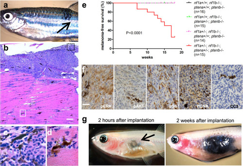
<italic>nf1a</italic><sup><italic>+/−</italic></sup><italic>;nf1b</italic><sup><italic>−/−</italic></sup><italic>;ptena</italic><sup><italic>+/−</italic></sup><italic>;ptenb</italic><sup><italic>−/−</italic></sup><italic>;p53</italic><sup><italic>M214K/M214K</italic></sup> zebrafish spontaneously develop melanomas with rapid growth.a Representative 16-week-old nf1a+/−;nf1b−/−;ptena+/−;ptenb−/−;p53M214K/M214K zebrafish with one spontaneous melanoma (indicated by arrow). b Hematoxylin and eosin (H&E) staining of the melanoma tumor shown in panel a (×5 magnification, scale bar = 200 μm). c Melanoma tumor cells from the black box in (b), magnified ×100. d Melanoma tumor cells that have invaded into the dorsal muscle from the white box in (b), magnified ×100. e Cumulative frequency of spontaneous melanomas arising in zebrafish with the indicated genotypes (generated by the inbreeding of the nf1a+/−;nf1b−/−;ptena+/−;ptenb−/−;p53M214K/M214K line, p < 0.0001, log-rank test). f Immunohistochemical analysis of melanoma tumor sections using antibodies to detect phosphorylated ERK1/2 (pERK), phosphorylated AKT (pAKT), phosphorylated S6 (pS6), proliferating cell nuclear antigen (PCNA) and cleaved caspase 3 (CC3) (×63 magnification, scale bar = 20 μm). The percentage of PCNA+ cells was determined by manually counting positive and negative melanoma cells in one representative high-power field (150–200 cells per field) within three independent tumor samples. g Pigmented nf1/pten-mutant melanoma cells were transplanted intraperitoneally into adult rag2−/− Casper zebrafish. The implanted melanoma cells (left panel, arrow) grew rapidly into secondary tumors (within 2 weeks; right panel).
|