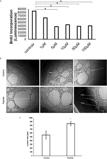
Angiogenesis in vitro analysis. (A) Cell proliferation assay in the presence of HS-binding peptide. Proliferation assays were performed using BrdU as described in Methods. HUVEC cells were treated with the HS-binding peptide at different doses for 18 hours. After treatment, BrdU was added into the cell culture and luminescence was analyzed. Assay performed in triplicate, the bar represents average and lines represent standard deviation. The proliferation is inhibited from 1 µM of HS-binding peptide and reaches maximum inhibition at 5 µM. *p < 0.05 (Kruskall-Wallis). (B) Tubular formation with endothelial cells. Control, Cells remain in the absence of treatment. Peptide, Cells were treated with the HS-binding peptide (10 µM) for 18 hours. The tubular formation was analyzed by microscopy 20x magnification. (C) Lumen size was measured, the bar represents average and lines represent standard deviation, *p < 0.05 (Kruskall-Wallis). Control, cells remain in the absence of treatment. Peptide, cells were treated with HS-binding peptide (10µM) for 18hours. It was verified that peptide was capable to inhibit the tubular formation of endothelial cells.
|
