
FIGURE
Fig 5
Fig 5
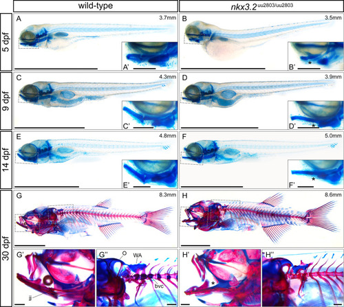
(A-F) lateral views of cartilage-stained wild-types and nkx3.2 mutants at 5, 9 and 14 dpf. (G-H) lateral views of cartilage- and bone-stained wild-types and nkx3.2 mutants at 30 dpf. Boxes in (A-H) indicate the zoomed-in regions in the insets or zoomed-in panels (G’, G”, H’, H”). The measurements in mm refer to standard length (SL). Asterisks indicate the fusion between Meckel’s cartilage and palatoquadrate in the jaw joint. jj–jaw joint, O–occipital region, bvc–basiventral cartilage, WA–Weberian apparatus. Scale bars: 2mm (A-H), 200μm (A’-H’ and G”-H”). |
Expression Data
Expression Detail
Antibody Labeling
Phenotype Data
| Fish: | |
|---|---|
| Observed In: | |
| Stage Range: | Day 5 to Days 30-44 |
Phenotype Detail
Acknowledgments
This image is the copyrighted work of the attributed author or publisher, and
ZFIN has permission only to display this image to its users.
Additional permissions should be obtained from the applicable author or publisher of the image.
Full text @ PLoS One