FIGURE

Figure 4

ID
ZDB-FIG-210813-8
Publication
Park et al., 2021 - Identification of de novo EP300 and PLAU variants in a patient with Rubinstein-Taybi syndrome-related arterial vasculopathy and skeletal anomaly
Other Figures
All Figure Page
Back to All Figure Page
Figure 4

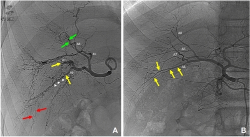
Comparison of visceral angiograms between the patient’s hepatic artery (A) and a normal hepatic artery (B). Overall, the “pruned branches” is a sign of normal peripheral hepatic arteries with extensive intrahepatic corkscrew and tortuous collateral arteries (A). Abrupt cut-off of second order branch of the hepatic artery types A6 and A7 (yellow arrows). Fine corkscrew collateral arteries along the A6 running course that replaces the normal A6 branch (white arrow heads). Intrahepatic tortuous collateral arteries developed in the peripheral liver (red arrows). Multiple stenosis and focal ectasia of the segmental hepatic artery (A8). This lead to the complete obstruction of the arteries and subsequent collateral development (green arrows). The normal A6 hepatic artery (B) showed typical smooth peripheral tapering out of the segmental hepatic artery (A6) with side-by-side ramification of the intrahepatic branches (yellow arrows).

Expression Data

Expression Detail
Antibody Labeling
Phenotype Data

Phenotype Detail
Acknowledgments
This image is the copyrighted work of the attributed author or publisher, and ZFIN has permission only to display this image to its users. Additional permissions should be obtained from the applicable author or publisher of the image. Full text @ Sci. Rep.