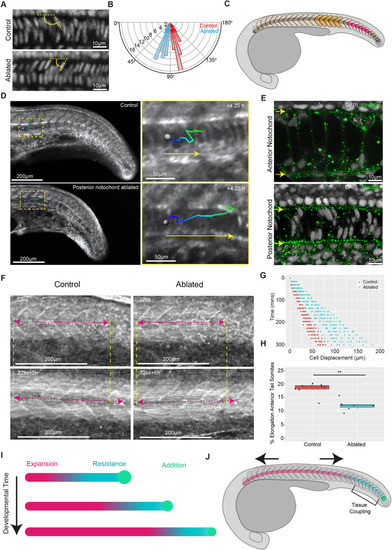
Notochord progenitors provide a source of resistance to notochord cell expansion, facilitating segmented tissue elongation in zebrafish embryos. (A) DAPI-stained notochord nuclei located in the anterior tail region in fixed control and ablated embryos. The angle between dorsally located nuclei and the notochord AP axis is indicated in yellow. (B) Polar histogram showing the distribution of angles between dorsally located nuclei and the notochord AP axis in control and ablated embryos (n=12 embryos per condition, five angles measured per embryo; P<0.001). (C) Schematic showing the ablated posterior unexpanded region (pink) and region used for measuring segmented-tissue elongation (orange). (D) Manual tracking (green-blue lines) of expanding notochord cells in a representative control and posteriorly-ablated embryo. Yellow arrows indicate direction of cell movement. (E) Vinculin and DAPI immunostaining in representative anterior and posterior notochord regions. Yellow arrows mark the dorsal (top) and ventral (bottom) extents of the notochord. (F) Elongation of a five-somite region in control and posteriorly ablated post-tailbud-stage embryos. The yellow dashed lines indicate the anterior and posterior extent of measured regions and the pink dashed arrows indicate the length of the measured region. (G) Notochord cell displacement over time in control and posteriorly ablated embryos (n=4 and n=5 embryos, respectively, 3 tracks per embryo). (H) Percentage elongation of a five-somite region in control and posteriorly ablated embryos (n=8 and n=7 embryos, respectively; **P<0.01, Mann–Whitney U test). (I) Notochord cell expansion progresses posteriorly along the axis. Expanding cells (pink) push against resisting unexpanded notochord cells (blue) added by posterior notochord progenitors (green), generating a stress along the notochord. (J) The somitic compartment anterior to the posterior resistance point undergoes an AP stretch (indicated by the black arrows). Tissue coupling between notochord cells and the somitic compartment is proposed to occur in the posterior of the embryo.
|
