FIGURE

Figure 3

ID
ZDB-FIG-210705-13
Publication
Follain et al., 2021 - Impairing flow-mediated endothelial remodeling reduces extravasation of tumor cells
Other Figures
All Figure Page
Back to All Figure Page
Figure 3

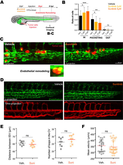
Inhibition of VEGFRs with sunitinib reduces endothelial remodeling in zebrafish embryos. (A) Experimental setup used: zebrafish are imaged at 3 hpi (hours post-injection). (B) Quantification of intravascular, remodeling and extravasated cells 3hpi. N cells: vehicle = 622, sunitinib 2 µM = 375, sunitinib 5 µM = 254. N embryos : vehicle = 62, sunitinib 2 µM = 34, sunitinib 5 µM = 27, Kruskal–Wallis test followed by Dunn's multiple Comparison test. (C) Representative image of arrested D2A1 cells in the caudal plexus of vehicle or sunitinib treated embryos. Vehicle: DMSO. (D) Representative confocal image of the caudal plexus architecture (upper panel) and blood flow (lower panel, RBC = red blood cells) 3 h post-treatment with sunitinib vs vehicle in Tg(fli1a:EGFP;gata1:DsRed). (E) Quantification of the effect of sunitinib on vessel architecture after 3 h of treatment with vehicle or 5 µM of sunitinib, N = 8, Student t test. (F) Blood flow perfusion profiles of the same embryos (from D) and quantification of the blood flow velocity in the caudal plexus of embryos in both conditions after 3 h of treatment with vehicle or 5 µM of sunitinib. N embryos: vehicle = 8, sunitinib 5 µM = 10. Mann Whitney test.

Expression Data

Expression Detail
Antibody Labeling
Phenotype Data

Phenotype Detail
Acknowledgments
This image is the copyrighted work of the attributed author or publisher, and ZFIN has permission only to display this image to its users. Additional permissions should be obtained from the applicable author or publisher of the image. Full text @ Sci. Rep.