
Figure 1
- ID
- ZDB-FIG-210630-41
- Publication
- Jerafi-Vider et al., 2021 - VEGFC/FLT4-induced cell-cycle arrest mediates sprouting and differentiation of venous and lymphatic endothelial cells
- Other Figures
- All Figure Page
- Back to All Figure Page
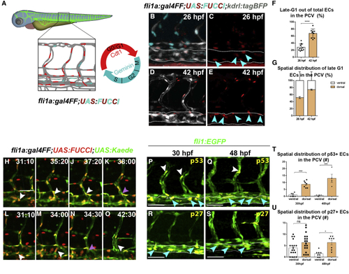
Endothelial cells sprout from the PCV in G1 phase (A) Schematic representation of cell cycle stages in ECs as highlighted by the (B–E) Selected confocal snapshots from a time-lapse series of a (F) Fraction of late-G1 ECs out of total PCV cells (n26 hpfembryos = 11; n42hpf embryos =9 ). Data show mean ± SEM (unpaired t test). (G) Spatial distribution of late-G1 ECs in the PCV at 26 and 42 hpf (n26 hpf embryos = 11; n42 hpf embryos =9 ). Data show mean ± SEM (unpaired t test). (H–O) Selected snapshots from a time-lapse series of a (P–S) Confocal images of 30 (P and R) and 48 (Q and S) hpf (T and U) Spatial distribution of p53+ (n30 hpf = 7, n48 hpf = 4) (T) and p27+ (n30 hpf = 18, n48 hpf = 8) (U) ECs in the PCV. Data show mean ± SEM (one-way ANOVA plus Tukey`s post-hoc test). Scale bars: 40 μm (B–E and H–O), 70 μm (P–S); ∗p < 0.05, ∗∗∗p < 0.001, ∗∗∗∗p < 0.0001; ns, not significant. |