Fig. 5
- ID
- ZDB-FIG-210517-14
- Publication
- Schoels et al., 2021 - Single-cell mRNA profiling reveals changes in solute carrier expression and suggests a metabolic switch during zebrafish pronephros development
- Other Figures
- All Figure Page
- Back to All Figure Page
|
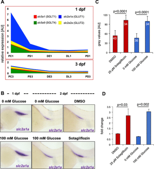
Rapid decline of glucose-transporting capacity during zebrafish pronephros development. A: glucose carriers of the glucose transporter (GLUT) and Na+-glucose transporter (SGLT) gene families rapidly declined after the first 24 h of development. Compared with 1 day postfertilization (dpf), very reduced expression of glucose transporters remained at 3 dpf. PC1, PS1, DE1, DL1, and PD1, proximal convoluted tubule, proximal straight tubule, distal early tubule, distal late tubule, and pronephric duct at 1 dpf; PC3, PS3, DE3, DL3, and PD3, proximal convoluted tubule, proximal straight tubule, distal early tubule, distal late tubule, and pronephric duct at 3 dpf. B: environmental glucose affects expression of glucose carriers. Although high-glucose concentrations (100 mM) had no effect on the expression of slc2a1 (GLUT1) at 1 dpf, glucose antagonized the downregulation of slc2a1a at 2 dpf. The SGLT inhibitor sotagliflozin also prevented downregulation of slc2a1a. C: quantification of the whole-mount in situ hybridization data shown in B. The numbers within the bars show the total number of analyzed embryos. The error bars indicate SDs. P values were calculated with a Student’s t test. D: quantitative RT-PCR for slc2a1a at 2 dpf confirmed the results from the whole-mount in situ hybridization quantification: high environmental glucose or sotagliflozin treatment maintained high levels of slc2a1a expression. P values were calculated with a Student’s t test. DE1, distal early tubule; DL1, distal late tubule; PC3; pronephric cells; PD1, pronephric duct; PS1, proximal straight tubule; SLCs, solute carriers. |

