|
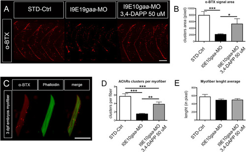
3,4-DAPP administration increased density and appropriate spatial positioning of AChRs. (A) Representative image of AChRs (α-BTX) in 4 somites of zebrafish embryos at 48 hpf collected during 3 distinct experiments. STD-Ctrl embryos: n = 32, untreated I9E10gaa-MO: n = 30, and I9E10gaa-MO treated with 3,4-DAPP 50 µM: n = 31, Scale bar = 25 µm. (B) Quantification of α-BTX-positive spots in the three experimental groups. (C) Representative image of single myofibers obtained after dissociation from 3 dpf embryos. Scale bar = 30 µm. (D,E) Quantification of AChR clusters in dissociated myofibers (D), and of myofiber average length (E) in the three experimental groups. Note that myofiber average lengths are comparable. Error bars in (B), (D), (E) are SEMs.
|