
Fig. 3
- ID
- ZDB-FIG-210316-9
- Publication
- Bertozzi et al., 2020 - Is zebrafish heart regeneration "complete"? Lineage-restricted cardiomyocytes proliferate to pre-injury numbers but some fail to differentiate in fibrotic hearts
- Other Figures
- All Figure Page
- Back to All Figure Page
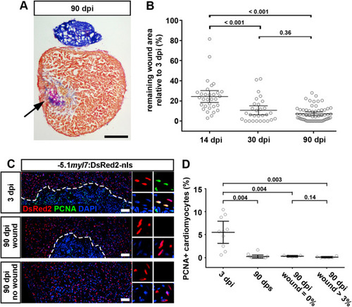
Fig. 3. Cardiomyocyte proliferation ceases in both scarred and unscarred hearts. (A) AFOG stained section at 90 dpi displaying incomplete scar resorption (arrow). Scale bar, 100 μm. (B) Average wound area does not significantly shrink between 30 and 90 dpi. n (hearts) = 31 (14 dpi), 29 (30 dpi), 58 (90 dpi). One-way ANOVA + Tukey’s multiple comparisons test. Observed difference 90 dpi vs 30 dpi = 3.5 (32% of 30 dpi wound area); smallest significant detectable difference = 5.8 (54% of 30 dpi wound area). (C, D) PCNA+ cardiomyocytes can be detected at 3 dpi, but not in 90 dpi hearts with or without wound. Scale bar, 100 μm n (hearts) = 10 (3 dpi), 12 (90 dps), 5 (90 dpi no wound), 6 (90 dpi wound). One-way ANOVA + Tukey’s multiple comparisons test. |
Reprinted from Developmental Biology, 471, Bertozzi, A., Wu, C.C., Nguyen, P.D., Vasudevarao, M.D., Mulaw, M.A., Koopman, C.D., de Boer, T.P., Bakkers, J., Weidinger, G., Is zebrafish heart regeneration "complete"? Lineage-restricted cardiomyocytes proliferate to pre-injury numbers but some fail to differentiate in fibrotic hearts, 106-118, Copyright (2020) with permission from Elsevier. Full text @ Dev. Biol.