
Fig. 4
- ID
- ZDB-FIG-210316-25
- Publication
- Jedrychowska et al., 2020 - Kcnb1 plays a role in development of the inner ear
- Other Figures
- All Figure Page
- Back to All Figure Page
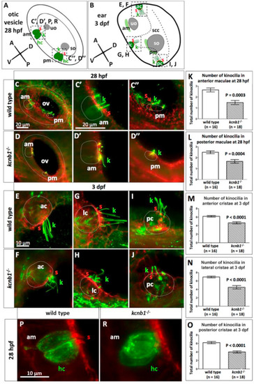
Fig. 4. The orientation of kcnb1−/− tether cell kinocilia is abnormal. (A, B) Schematic representation of the otic vesicle at 28 hpf (A) and ear at 3 dpf (B). (C, D) Lateral view of the otic vesicles at 28 hpf. (C′, D′, C″, D″) Magnification of anterior maculae (C′, D′) and posterior maculae (C″, D″). kcnb1−/− kinocilia (green) are oriented abnormally. (K, L) Number of kcnb1−/− kinocilia in anterior and posterior maculae at 28 hpf was lower compared with wildtype control. (E, F) Magnification of anterior cristae at 3 dpf, (G, H) Lateral cristae at 3 dpf, (I, J) Posterior cristae at 3 dpf. (F, H, J) kcnb1−/− kinocilia formed abnormally oriented “looped strings”. (M–O) At 3 dpf, the number of kinocilia in anterior, lateral and posterior cristae was lower compared with wildtype controls. (P–R) In mutants, hair cells were present, but stereocilia were not detected. Imaging was performed using light-sheet fluorescence microscopy with a 63 objective (C, D00) and 40 objective (E-J, P-R). Staining: green (anti-acetylated tubulin), red (phalloidin) in (C-J), green (antiotoferlin) and red (phalloidin) in (P-R). Data are expressed as mean SEM (unpaired Student’s t-test). ac, anterior crista; am, anterior macula; dpf, days post fertilization; hc, hair cell; hpf, hours post fertilization; k, kinocilium; lc, lateral crista; ov, otic vesicle; pc, posterior crista; pm, posterior macula; s, stereocilia; scc, semicircular canals; so, saccular otolith; uo, utricular otolith. |
Reprinted from Developmental Biology, 471, Jedrychowska, J., Gasanov, E.V., Korzh, V., Kcnb1 plays a role in development of the inner ear, 65-75, Copyright (2020) with permission from Elsevier. Full text @ Dev. Biol.