
Fig. 3
- ID
- ZDB-FIG-210316-24
- Publication
- Jedrychowska et al., 2020 - Kcnb1 plays a role in development of the inner ear
- Other Figures
- All Figure Page
- Back to All Figure Page
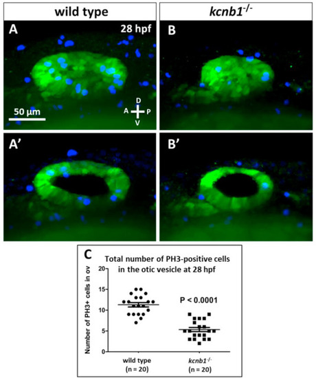
Fig. 3. Cell proliferation is low in the kcnb1−/− otic vesicle at 28 hpf . (A, B) Maximum intensity projections of z-stacks that represent the otic vesicle at 28 hpf in the wildtype controls (A) and kcnb1−/− mutant (B). (A′, B′) Optical cross-sections of the otic vesicles. (C) Total number of PH3-positive cells in the mutant’s otic vesicles was low at 28 hpf. Imaging was performend using light-sheet fluorescence microscopy (40 objective). Staining: green (anti-GFP), blue (anti-phospho-histone 3). The ET33-mi2A zebrafish line was used as a transgenic background. The data are expressed as mean SEM (unpaired Student’s t-test). |
Reprinted from Developmental Biology, 471, Jedrychowska, J., Gasanov, E.V., Korzh, V., Kcnb1 plays a role in development of the inner ear, 65-75, Copyright (2020) with permission from Elsevier. Full text @ Dev. Biol.