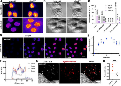
In cellulo uncaging of pcHoechst. A) Epifluorescence imaging of live HeLa cells incubated with Hoechst33342 or pcHoechst (10 μM each) before and after 4 s of UV light irradiation shows an increase in nuclear fluorescence for pcHoechst‐treated cells. Scale bar: 20 μm. B) As in (A), but phase contrast images. C) Cells from (A) were imaged over 24 h to determine single cell viability to progress through a complete cell cycle during compound and UV treatment, numbers indicate absolute cell division events. D) Confocal images of live HeLa cells incubated with pcHoechst (10 μM). Between each image, UV light was applied with higher intensity to uncage pcHoechst. Scale bar: 20 μm. E) Fluorescence increase over time upon pcHoechst uncaging (n=15 cells). F) Line scans result in fluorescence increase over time upon pcHoechst uncaging (from dashed line in D). G) Colocalization of cytosolic signals from pcHoechst with the lysosomal system labeled with LysoTracker Red. Scale bar: 5 μm. H) Pearson's correlation R value for cytosolic colocalization is positive for co‐applied pcHoechst and LysoTracker Red, and negative in controls, thus arguing for extranuclear signals stemming from acidic compartments; n=6 cells.
|
