|
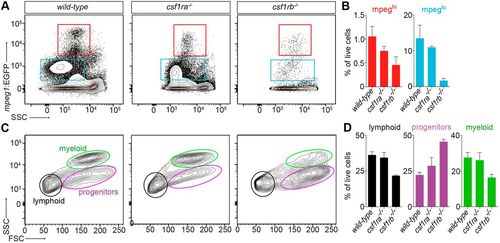
Adult zebrafish csf1rb mutant display hematopoietic deficiencies. (A,B) Flow cytometry analysis of WKM cell suspensions from wild-type, csf1ra−/− and csf1rb−/− adult fish carrying the mpeg1:EGFP reporter. (A) The mpeg1:EGFPhi fractions identify mature macrophages (red frames), whereas the mpeg1:EGFP lo fractions contain mainly IgM-expressing B lymphocytes (blue frames) (Ferrero et al., 2020). (B) Percentage of mpeg1hi and mpeg1lo cells for each genotype, relative to the whole living WKM population. Each bar represents the mean±s.e.m. for three individuals. (C) Scatter profiles of WKM in typical wild-type (left panel), csf1ra−/− (middle panel) and csf1rb−/− (right panel) adult fish. (D) Percentage of cells in the lymphoid (black), myeloid (green) and progenitor (purple) fractions, relative to the whole hematopoietic cell population. Each bar represents the mean±s.e.m. for three individuals.
|