
FIGURE
Figure 3
- ID
- ZDB-FIG-210227-8
- Publication
- Yang et al., 2021 - Ca2+-Dependent Glucose Transport in Skeletal Muscle by Diphlorethohydroxycarmalol, an Alga Phlorotannin: In Vitro and In Vivo Study
- Other Figures
- All Figure Page
- Back to All Figure Page
Figure 3
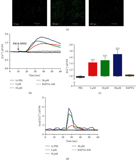
Detection of cytosolic Ca2+ levels in the absence of extracellular Ca2+ using the Fluo-4 indicator in the myotubes. (a) Representative images of myotubes at time zero (0 sec) and after stimulation with 30 μM of DPHC at time 30 sec and 60 sec. (b) Traces and (c) box plot representation of Ca2+ levels in response to addition of DPHC or BAPTA-AM in myotubes. The fluorescence levels of single myotubes after addition of DPHC or BAPTA-AM was also monitored (d). *p<0.05 and **p<0.001 compared with the PSS group. |
Expression Data
Expression Detail
Antibody Labeling
Phenotype Data
Phenotype Detail
Acknowledgments
This image is the copyrighted work of the attributed author or publisher, and
ZFIN has permission only to display this image to its users.
Additional permissions should be obtained from the applicable author or publisher of the image.
Full text @ Oxid Med Cell Longev