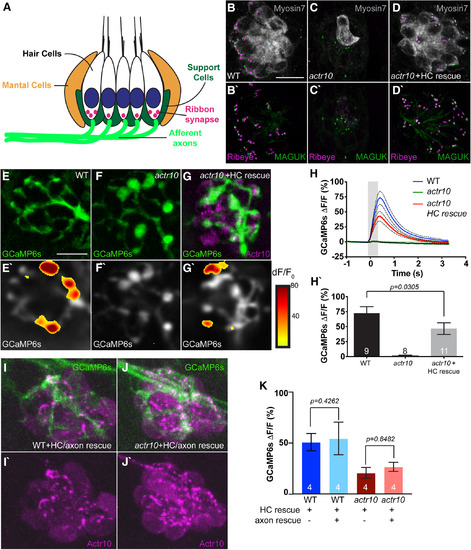
Postsynaptic axon response to stimulation is unaffected by mitochondrial health. A, Schematic of the neural circuit in a sensory neuromast of the pLL. Apical stereocilia on HCs are deflected by water movement and signal through ribbon synapses to afferent (postsynaptic) axons. Support cells surround HCs. B, C, actr10nl15 mutants have fewer sensory HCs as assayed by Myosin VIIa immunolabeling. D, D', HC and synapse number is rescued by expressing RFP-tagged Actr10 in HCs (HC rescue) using the Tg(myo6b:mRFP-actr10)y610 transgenic. Ribeye: presynapse; MAGUK: postsynapse. E–G, GCaMP6s expression in an axon terminal of a WT (E), actr10nl15 mutant (F), and actr10nl15 mutant with HC rescue (G) at 5 dpf. E'–G', Shown are spatial patterns of GCaMP6s signal increases in afferent terminals during stimulation. GCaMP6s signals are colorized according to the dF/F0 heat maps and are superimposed onto a prestimulus baseline GCaMP6s image. H, H', The average change in GCaMP6s fluorescence intensity on stimulation, shown in plots (H) and quantification (H': WT: 73.26 ± 10.62; actr10nl15: 1.53 ± 0.76; actr10nl15+HC rescue: 43 ± 7.78), revealed a reduction in actr10nl15 mutants with and without HC rescue (ANOVA). Black dotted lines represent SEM. I, J, Actr10 rescue in axons using mosaic expression of the 5kbneurod:mRFP-actr10 transgene (axon rescue) in the background of the Tg(myo6b:mRFP-actr10 y610 transgenic (HC rescue). K, Rescuing Actr10 in neurons does not rescue postsynaptic axonal activity based on average GCaMP6s fluorescence (t test). WT: 50.90 ± 8.45; WT+HC rescue: 54.52 ± 16.0; actr10nl15: 20.69 ± 5.37; actr10nl15+HC rescue: 26.65 ± 4.45. Sample sizes indicated on graph. Scale bar: 10 µm. All data are mean ± SEM.
|
