
Fig. 6
- ID
- ZDB-FIG-210217-84
- Publication
- Shao et al., 2020 - Serum lipoprotein-derived fatty acids regulate hypoxia-inducible factor
- Other Figures
- All Figure Page
- Back to All Figure Page
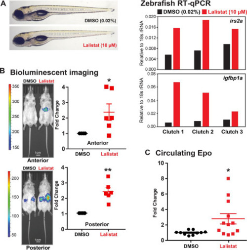
Figure 6. LAL inhibition activates HIFα in animals. A, representative images of WT zebrafish larvae (5 days postfertilization) treated with lalistat (10 μm) or vehicle control DMSO (0.02%) for 2 h are shown. Expression of HIFα targets irs2a and igfbp1a (relative to 18s rRNA, RNA pooled from five larvae) was analyzed using RT-qPCR. B, ODD-Luc mice received subcutaneous injection of either DMSO or lalistat (20 mg/kg) three times per week for 2 weeks. Shown are representative bioluminescent images (captured 2 min after luciferin injection) of ODD-Luc mice treated as indicated. Bioluminescent signals from lalistat-treated mice were normalized to vehicle control (n = 6, mean ± S.E. (error bars)). p values from a single-column t test (Lalistat versus DMSO) are shown: *, p < 0.05; **, p < 0.005. C, circulating Epo levels from lalistat-treated mice (n = 12, mean ± S.E.) were normalized to vehicle control (n = 10, mean ± S.E.). p values from a single-column t test (lalistat versus DMSO) are shown: *, p < 0.05. |