
Fig. 1
- ID
- ZDB-FIG-210217-79
- Publication
- Shao et al., 2020 - Serum lipoprotein-derived fatty acids regulate hypoxia-inducible factor
- Other Figures
- All Figure Page
- Back to All Figure Page
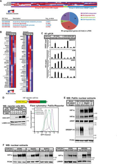
Figure 1. Lipoprotein depletion activates HIFα under normoxia. A, patient-derived human PDAC cell line Pa03c (WT), SCAP KO cells (S), and HIF1A HIF2A DKO cells (H) were cultured in FBS or LPDS for 16 h. Gene expression was determined using Illumina bead arrays. LPDS-induced genes (≥2-fold) were analyzed for GO term enrichment using GOrilla/REVIGO. GO terms related to the SREBP or HIF pathway are highlighted in blue or red, respectively. A clustered heatmap of LPDS-induced genes was generated by GenePattern 2.0. B, a clustered heatmap of 99 genes induced upon lipoprotein depletion (≥2-fold) was generated by GenePattern. Group A, induction in LPDS required SCAP, but not HIFα. Group B, induction in LPDS required HIFα, but not SCAP. Boldface, underlined genes are known transcriptional targets of SREBP or HIF in Group A or B, respectively. C, WT or HIF1A HIF2A DKO Pa03c cells were cultured for 16 h in FBS in the presence of DMSO (0.1%) under normoxic (FBS) or hypoxic (1% O2) conditions or in LPDS in the presence of DMSO (0.1%) (LPDS) or Site-1 protease inhibitor PF-429242 (50 μm) (L+PF) to inhibit SREBP. Gene expression for SREBP or HIF transcriptional targets measured by RT-qPCR was normalized to vehicle-treated Pa03c cells cultured in FBS. Error bars, S.E. of -fold changes from three biological replicates (mean ± S.E.). D, diagram of HIF reporter cell line that is a Pa03c clone stably expressing HIF1α ODD-d2EGFP under the control of five tandem HREs. Shown are immunoblots (WB) of whole-cell lysates or flow cytometry analysis from parental Pa03c cells cultured for 24 h in FBS and HIF reporter cells cultured for 24 h in FBS, LPDS, or FBS with DMOG (1 mm), a cell-permeable prolyl-4-hydroxylase inhibitor. E, immunoblots of nuclear extracts from Pa03c cells cultured for 16 h in FBS in the presence of DMSO (0.1%) under normoxic or hypoxic (1% O2) conditions or in LPDS in the presence of DMSO (0.1%) or site-1 protease inhibitor PF-429242 (50 μm) to inhibit SREBP. PCNA served as a loading control. F, immunoblots of nuclear extracts from the indicated cell lines cultured for 16 h in FBS, LPDS, or FBS in 1% O2. PCNA or LMNB1 served as a loading control. ACHN, renal cell adenocarcinoma; HepG2, hepatocellular carcinoma; BxPC3, pancreatic adenocarcinoma; U2OS, osteosarcoma; HEK293, embryonic kidney. |