
Fig. 5
- ID
- ZDB-FIG-210216-73
- Publication
- Rieckhoff et al., 2020 - Spindle Scaling Is Governed by Cell Boundary Regulation of Microtubule Nucleation
- Other Figures
- All Figure Page
- Back to All Figure Page
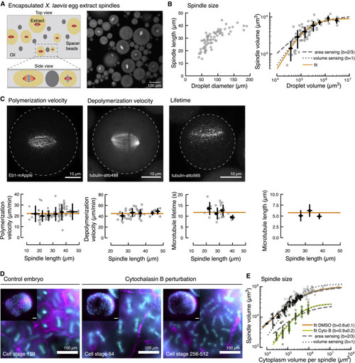
Figure 5. Spindle Scaling in Encapsulated Xenopus Egg Extracts and Cytokinesis-Inhibited Zebrafish Embryos (A) Spindle assembly in cell-like compartments by encapsulating Xenopus laevis egg extract in inert oil. Droplets are compressed between spacer beads to improve image quality and spindle orientation. Microtubules are labeled with tubulin-atto488. (B) Spindle scaling with droplet size is consistent with a simple limiting component model in the absence of membrane partitioning. Left: spindle length in encapsulated Xenopus laevis egg extract scales with droplet diameter. Each dot denotes an individual measurement (n = 77). Right: spindle volume scales linearly with droplet volume (gray dotted line and fit in orange). (C) Microtubule dynamics remain constant over a large range of spindle sizes in encapsulated Xenopus laevis egg extract (see also Video S6). Altogether these measurements determine the average microtubule length in the spindle (right). Gray dots denote individual measurements (n= 32, 48, and 27), and black dots are averages over 5 μm spindle size intervals. Error bars show SDs. The orange lines show the average values. (D) Pharmacological inhibition of cytokinesis using cytochalasin B in zebrafish embryos prevents the formation of new cell boundaries leading to spindles sharing the same cytoplasm. Left: a control embryo at cell stage 128 shows normal cellularization (spindles in cyan and cell boundary in magenta). Right: a perturbed zebrafish embryo at two distinct cell stages showing that new boundaries are not formed (cytochalasin B was added at cell stage 8), but spindles continue to divide and scale as they share the embryo cytoplasm. (E) Spindle scaling in control DMSO condition scales as the cell boundary (dark gray points, n = 41) whereas in the cytochalasin B perturbation, spindles scale linearly with the volume per spindle (green points, n = 69). The overall amplitude of spindle volumes is additionally decreased in the cytochalasin B condition, consistent with surface sequestration of a microtubule nucleation inhibitor. As reference, data from embryos without DMSO or cytochalasin B treatment is shown in light gray. |