
Fig. 2
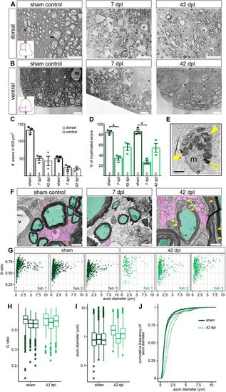
SCI results in axonal and myelin sheath loss, and different myelin patterns between individual fish. (A,B) Myelinated and non-myelinated axons are decreased after SCI. Overview images are TEM images of transverse sections. D, dorsal; V, ventral (m, Mauthner axon; v, blood vessel). Scale bars: 20 µm. (C) Quantification of axons in a 500 µm2 area after SCI. Data are mean±s.e.m. Numbers in the plots indicate the number of experimental animals. (D) Percentage of myelinated axons after SCI. Data are mean±s.e.m. Numbers in the plots indicate the number of experimental animals. *P≤0.05 (Kruskal–Wallis followed by Dunn's multiple comparisons post-hoc test). Significance is shown compared to sham control. (E) Macrophage/microglia engulfing myelin debris (filled yellow arrowheads) and lipid droplets (empty yellow arrowhead). Scale bar: 1 μm. (F) SCI results in thinner/still forming myelin sheaths after (re-)myelination. Axons are pseudo-coloured (green, myelinated; magenta, non-myelinated; yellow arrowheads, newly formed myelin sheaths; v, blood vessel). Images are TEM close-ups of transverse sections. Scale bars: 1 µm. (G) G-ratio over axon diameter for all individual fish investigated from the sham control and the 42 dpl group. Each dot represents an axon. (H) G-ratio analysis of all individual fish investigated from the sham control (dark green) and 42 dpl group (light green). Data are presented as box-and-whisker plots, each box presents one of the individual fish shown in G (150-1524 axons were measured per fish). (I) Axon diameter of all individual fish investigated from sham control and 42 dpl group. Data are presented as in H. (J) Cumulative frequency of axon diameter of myelinated axons in sham controls and at 42 dpl. |