
Fig. 5
- ID
- ZDB-FIG-210203-13
- Publication
- Huang et al., 2020 - GFP expression pattern in pituitary and gonads under the control of nuclear progesterone receptor promoter in transgenic zebrafish
- Other Figures
- All Figure Page
- Back to All Figure Page
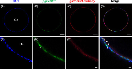
Cellular localization of green fluorescent protein (GFP) under the control of pgr promoter in ovarian follicles layer in Tg(pgr:egfp/gsdf:nfsB‐mCherry). The ovarian follicle consisted of an oocyte and a follicle cell layer (A, A′) which showed both GFP (B, B′) and mCherry (C, C′) signals. Most of GFP‐positive cells expressed mCherry signals (D), and located at inner follicle layer (D′). A few of GFP‐positive cells without mCherry signals showed strong GFP signals (B′, D′) and located at outer follicle layer (D′). The blue fluorescence represented the nucleus which is stained by Hoechst33342. Oc, oocyte; white arrow, thecal cell; white arrowhead, granulosa cell. Scale bars = 10 μm. Pgr, progesterone receptor |